CPRIT Imaging Core
| Home | Publications | Rates | Reservations |
|---|

The mission of the Amarillo Imaging Core is to provide researchers across West Texas and beyond access to cutting edge instruments and imaging service, covering the range from small animal live imaging to optical super resolution microscopy.
CPRIT Imaging Core Instruments:
About:
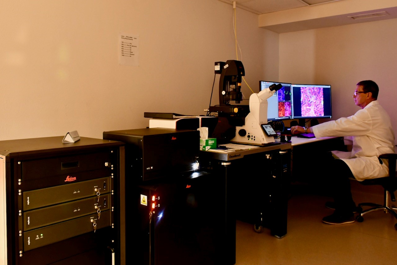
Spectral confocal microscope with optical super resolution capability (Stimulated Emission Depletion, STED) enabling resolution down to ~30 nm. FAst Lifetime CONtrast (FALCON) maps fluorescence lifetime, enhancing contrast in combination with STED (tau-STED). The system is further equipped with an optional on-stage live cell imaging chamber (Okolab) and an AI-based quantitative image analysis package (AIVIA).
Key Features:
- DMi8 inverted microscope stand
- Immersion lenses for super resolution imaging (< 50 nm)86x (water), 93x (glycerol), 100x (oil)
- White light laser, 440nm – 790 nm, 405 nm UV laser
- Stimulated Emission Depletion (STED) module for optical super resolution
- STED lasers 592 nm, 660 nm, 775 nm
- 4 hybrid detectors, resonant scanner, transmitted light detector
- Fluorescence lifetime imaging (FALCON), tau STED.
- On-stage live cell incubation system (Okolab) for normoxia, hypoxia, or hyperoxia
- LAS X Control and Analysis Software
- Deconvolution (Lightning Expert)
- AIVIA 3D suite
About:
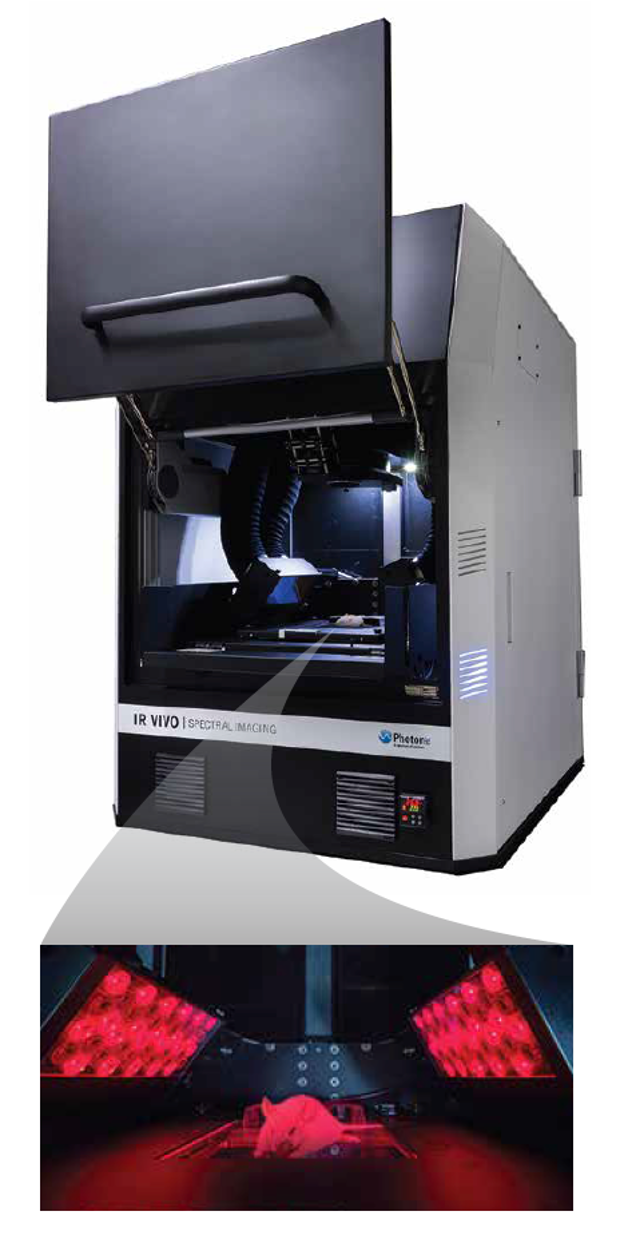
Unique small animal imager that it operates at near infra-red (INR) to infrared (IR) wavelengths and avoids the various complications associated with absorption scattering and the autofluorescence of light in living tissues. Using the first (NIR) and second biological window (NIR-II) from 900-1620 nm, the IR VIVO provides multispectral or hyperspectral infrared imaging capabilities for in vivo studies on small animals including rats.
About:
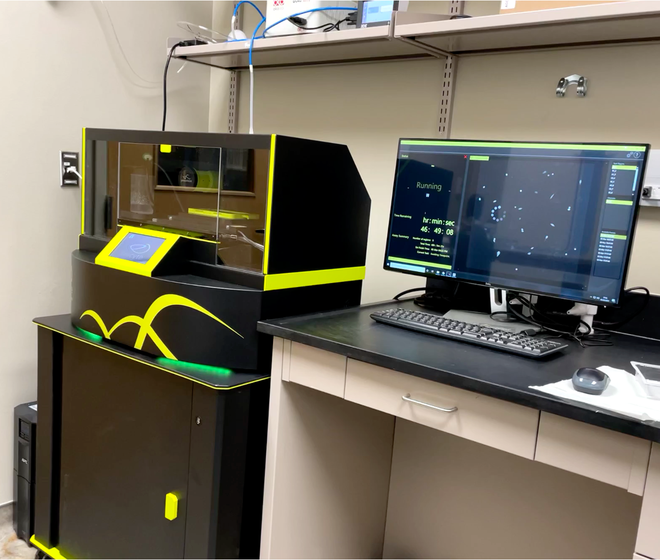
The Livecyte Kinetic Cytometer is a complete imaging and analysis system optimized for label-free long term, non-invasive monitoring of live cells. It utilizes Ptychographic Quantitative Phase Imaging (QPI), fluorescence and bright field imaging. Ptychographic QPI allows robust automatic tracking and behavioral analysis of a significant number of individual cells within heterogeneous cell populations without labels. Unique morphological, temporal, and dynamic phenotypic data make it simple to gain behavioral information of individual cells of cell clusters through live cell imaging. Achieve more efficient imaging by using every well in any plate, up to 96 well format.
About:
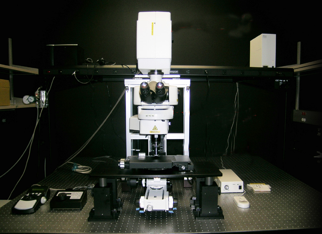
In live cells and tissue imaging, a key technology is two photon laser scanning microscopy (TPLSM). It minimizes phototoxic effects and offers the possibility to image deep into tissues.
Key Features:
- Simultaneous excitation imaging with two-wavelength IR laser
- Deep in vivo imaging with super high-sensitive GaAsP NDD
- Ultrahigh-speed imaging of up to 420 frames per second
- Multi-color in vivo imaging
- Deep specimen imaging with non-descanned detectors (NDD)
- Nikon's high-NA objectives are ideal for multiphoton imaging
- Auto laser alignment when changing multiphoton excitation wavelength
- Two types of scanning head enable high-speed, high-quality imaging
- NIS-Elements C acquisition and analysis software
Note About Citations: Please acknowledge the CPRIT Imaging Core in all publications or grant proposals as follows: "...funded by the Core Facility Support Award (grant number RP200572) from the Cancer Prevention and Research Institute of Texas (CPRIT) to the Imaging Core, Texas Tech University Health Sciences Center at Amarillo."
Contact Us
 |
Ulrich Bickel, M.D.Associate Dean of Sciences |
 |
Research Lab Manager |
 |
Zijuan "Amy" Liu, Ph.D.Research Lab Manager |
|
